Cells we were taught to ignore have been caught on tape doing the unthinkable.
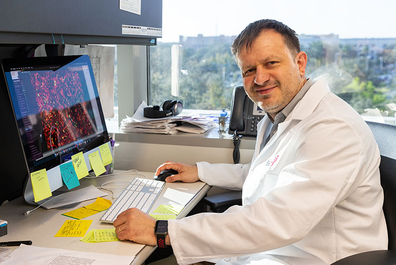
Sometime around 2009, Andriy Marusyk was staring down the microscope in the lab at the Dana-Farber Cancer Institute in Boston, checking in on his latest experiment aimed at understanding how cancerous and healthy cells interact together in tumors, when he made a provocative discovery. Cancer cells were having sex.
The experimental setup was straightforward, if sophisticated. First mix different populations of cells together, label each with different colored fluorescent tags—red for the cancer cells, green for the healthy ones—and then watch as they go about their daily lives. Slowly he scanned across the plastic dish, monitoring the cells as they passed under the lens. Red. Green. Green. Red. Red. Green. Then… orange?
His first reaction was to ignore it and move on. Strange things can happen in the world of lab-grown cells, and this orange blob was more than likely to be an irrelevant artifact. But still he kept coming across unusually large orange cells carrying both fluorescent markers.
His first thought was that two cells must have fused together to create some kind of hybrid, so he started looking more closely for evidence of cell fusions. Were the orange cells larger than the others? Did they have more than the regular amount of DNA? The more he looked the more he found evidence of cancer cells fusing with healthy cells or with each other. Not many, but enough to make him think there was something weird going on.
When he started asking around, colleagues told him they’d seen something similar, but it didn’t mean anything.
I first heard of Marusyk’s discovery while researching my latest book, Rebel Cell: Cancer, Evolution and the New Science of Life’s Oldest Betrayal. During those early stages of the writing process, I would occasionally hear hints of something so transgressively bizarre that it made my head spin.
Cell fusions happen all the time in the body and in the lab. For years biologists have studied their occurrence during the formation of osteoclasts—large cells that break down and remodel bone tissues—in the placenta in early pregnancy, or during infection with certain viruses like HIV. Spotting this process occurring in the unusual environment of a laboratory dish isn’t so strange.
Unusually large cells have also been observed in cancer for more than 150 years, going back to the early German pathologist Rudolf Virchow, who diligently sketched the “curious bodies, provided with large nuclei and nucleoli, which are described as the specific, polymorphous cells of cancer” that he noticed within a tumor. Taking this idea further, in 1911 his fellow countryman Otto Aichel proposed that cancer might spread through the body through tumor cells fusing with immune cells. Even though Aichel was wrong in the details, his concept wasn’t far off.
Cancer cells have also been caught completely engulfing healthy cells (a phenomenon known as emperipolesis), and fusions between cancer and immune cells have been found in several tumor types, including pancreatic cancer and melanoma. However, these fusions were thought to be terminal—the resulting, freakish hybrid cells were believed to be unable to grow and destined to die.
While plenty of oncology researchers staring at tumor sections through their microscopes saw these Frankenstein fused cancer cells through the years, most of them were not interested. They simply adjusted their microscopes and turned their focus elsewhere.
Marusyk couldn’t move on. He was stuck wondering why these cell fusions kept occurring and what role they could be playing in the cancer process. In 2015 he left Dana-Farber to set up his own lab at the Moffitt Cancer Center in Tampa, Florida, hiring a young Ukrainian researcher, Daria Miroshnychenko, to start working on the problem.

Caught in the act
One of the first challenges was developing a way to accurately detect and measure the extent of cell fusion events. Scanning through millions of cells with a microscope was too time consuming and inaccurate, while automated cell sorting or DNA sequencing techniques actively discarded ambiguous or mixed data resulting from exactly the kind of cell fusion events Marusyk and Miroshnychenko were looking for.
They were using a new technique called image-based cytometry, which whizzes thousands of cells past a tiny camera, snapping a photo of each one as it goes. Miroshnychenko found a small but significant proportion of cell fusions after growing different types of cancer cells together with tumor-associated fibroblast cells for three days. One thing they noticed right away was that some of the fused cells were far more active than might have been expected.
While many couldn’t divide, a small number of fused cells were able to proliferate, both in the lab and when transplanted into animals. In some cases, the cell fusions divided faster and their offspring became more invasive than the original cancer cells from which they came, suggesting that they could be playing a role in the development of aggressive tumors in real-life situations.
Are advances in technology finally allowing researchers to catch cancer cells in the act?
The product of many of these cell fusions were bulky, swollen cells, known as polyploids, which have twice the regular amount of DNA. But curiously, Miroshnychenko discovered that some of them had dropped back down to a normal amount of DNA after dividing. When she and her colleagues looked closely at the cells’ genomes, they found evidence of so-called parasexual recombination, where the fused cells were swapping bits of DNA around before dividing, creating new “daughter” cells with genetic properties that were a mix of “mom” and “pop.”
Was this, at long last, the smoking-gun hard evidence of cancer cell sex? If so, it brought with it major implications for our understanding of how tumors evolve within the body and become resistant to therapy.
This would be huge, if true. Our current model of how cancers pick up mutations and evolve within the body relies on the assumption that cancer cells reproduce asexually, without the ability to swap genetic tips and tricks between themselves. Yet beyond a handful of intriguing but low-profile papers, there has been little hard evidence for these cellular hookups.
As Marusyk explains, most techniques for studying cancer cells, such as DNA sequencing or flow cytometry, actively discard any evidence of cell fusions as being errors or artifacts, therefore many come to the conclusion that cell fusion doesn’t exist.
“This reflects a lack of understanding in how these techniques work,” he argues. “Absence of evidence is not evidence of absence, and there’s no way that the sequencing pipeline can detect cell fusions, as it’s built into the algorithm to reject them.”
At long last, are advances in technology finally allowing researchers to catch cancer cells in the act?
Pooling deadly resources
The entire field of cancer research and treatment is built on the idea that tumor cells can only reproduce by splitting in two. This means that any traits arising in a cancer cell due to a new genetic mutation, such as the ability to migrate around the body, invade new tissues, or resist the effects of treatment, must be restricted to that one lineage of cancer cells.
But if it’s possible for cancer cells to fuse together, pooling their genetic assets and then dividing to create even more deadly offspring, then this overturns what we think we know about how tumors grow and evolve within the body.
Parasexual recombination doesn’t introduce new mutations into a cell’s DNA. Instead, it allows cells with different mutations to recombine and reshuffle their respective genetic cards to create new combinations. This fits neatly with the emerging view of cancer as a Darwinian evolutionary process, with tumor cells diversifying and picking up new genetic changes that may enable them to better survive within the environment of the body.
This ability to evolve becomes even more important when the environments in which the cancer cells find themselves change, for example with the application of treatments like chemotherapy or radiotherapy, or under conditions of reduced oxygen or nutrients, which are often found within tumors.
Teaming up with computational biologist David Basanta at the Moffitt, Miroshnychenko and Marusyk created a mathematical model to show that this parasexual activity was capable of generating the kinds of extreme genetic diversity seen in advanced metastatic cancers, which has profound implications on the evolutionary capabilities of the cells.
“When cancer cells can have ‘sex’, then you can generate more diversity and create new combinations of mutations that previously existed in separate lineages,” Marusyk says. “So in order to understand cancer evolution and resistance to therapy, you have to account for the possibility that there is at least some occurrence of cell fusions.”
Figuring out the frequency and significance of these cell fusions will be important, Marusyk explains, because if they tend to occur between cells that are very similar to each other, then we can probably ignore it. On the other hand, in triple negative breast cancers, genetic heterogeneity is a defining feature. “So you are more likely to have a fusion between genetically dissimilar cells that would have a much larger evolutionary impact,” he says.
Mysterious giants and accelerated evolution
The idea that cancer cell fusions might be playing an important role in the emergence of resistance to treatment was neatly demonstrated in recent work by Kenneth Pienta and Sarah Amend at Johns Hopkins University in Baltimore, which also started from a curious observation down a microscope.
While looking at prostate cancer cells growing in the lab, Amend would occasionally spot large polyploid cells with at least double the normal amount of DNA. That’s not necessarily unusual: Cancer cells can get stuck at the point where they’ve copied their DNA but are unable to split into two new cells, so they’re normally overlooked and ignored by researchers. Yet like Marusyk, Amend and Pienta felt like there must be something more to these mysterious giants.
To investigate further, they turned to a device nicknamed the Evolution Accelerator—a hexagonal microfluidic chip no bigger than a fingernail, which was originally designed as a way of studying the emergence of antibiotic resistance in bacterial cells. Similar to the arena in The Hunger Games, the Evolution Accelerator is a miniature landscape in which cancer cells compete with each other, created from tiny chambers linked by tunnels that are small enough for regular cancer cells to move through but not the larger polyploid cells.
“Realizing the importance of such a critical cell type that we’re usually taught to ignore for so long was frankly startling.”
Unlike cells growing in a flat plastic Petri dish, which all experience the same conditions, the structure of the Evolution Accelerator meant that Amend and her colleagues could set up different zones across the chip, ranging from high concentrations of the common prostate cancer drug docetaxel on one side to low levels on the other. Then they added drug-sensitive prostate cancer cells into the arena and watched what happened.
Using time-lapse microscopy, the researchers followed the cells over several weeks as they explored their silicon setting. Strikingly, more and more giant polyploid cells quickly began to appear, especially in the areas with the highest levels of docetaxel, suggesting they had evolved resistance to the treatment.
Looking more closely at the footage, Amend was stunned to see that many of these polyploid cells were being created through cell fusions, rather than failures of cell division as expected. And she was even more shocked as she watched these giant cells split back into smaller “daughter” cells, all of which were now also resistant to the drug.
“It was a surprise observation, but we paid attention to it,” Amend says. “As a trained cell biologist, realizing the importance of such a critical cell type that we’re usually taught to ignore for so long was frankly startling.”
The results from Marusyk and Amend’s teams are an important addition to the steadily growing pile of evidence to support the importance of fusion and polyploid cells in cancer for generating evolutionary diversity and resistance to treatment. Although it’s technically much harder to spot parasexual cancer cell antics in real-life tumors, Amend is convinced that the more we look, the more we will find.
“If you know what to look for you can find them,” Amend explains. “Once you’re aware that they’re there, you can just look at images of tumors, and they are present. Once you see what these cells and their progeny are able to do, it really clarifies that this is the cell state we need to understand and target if we are to have any hope of eliminating cancer.”
Her own moment of clarity came while watching a movie of the Evolution Accelerator in action, as cell after cancer cell mated, split, and acquired resistance in real time.
“I’ve seen it hundreds of times,” she says, “and I still get goosebumps because it’s that terrifying.”


